| CAS NO: | 21561-09-1 |
| 规格: | ≥98% |
| 包装 | 价格(元) |
| 5mg | 电议 |
| 10mg | 电议 |
| 25mg | 电议 |
| 50mg | 电议 |
| 100mg | 电议 |
| 250mg | 电议 |
| 500mg | 电议 |
| Molecular Weight (MW) | 281.32 |
|---|---|
| Formula | C16H15N3O2 |
| CAS No. | 21561-09-1 |
| Storage | -20℃ for 3 years in powder form |
| -80℃ for 2 years in solvent | |
| Solubility (In vitro) | DMSO:>10mM |
| Water: | |
| Ethanol: | |
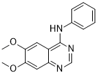
| Chemical Name | 6,7-Dimethoxy-N-phenylquinazolin-4-amine |
| Synonyms | WHI-P258; WHI-P 258; WHI-P258; WHIP258 |
| SMILES Code | COC1=CC2=NC=NC(NC3=CC=CC=C3)=C2C=C1OC |
| In Vitro | In vitro activity: WHI-P258 is a potent and selective Janus kinase 3 (JAK3) inhibitor discovered from homology modeling. Potent and specific inhibitors of JAK3 such as WHI-P131 may provide the basis for the design of new treatment strategies against acute lymphoblastic leukemia, the most common form of childhood cancer. WHI-P258 was identified from a novel homology model of the kinase domain of Janus kinase (JAK) 3 used for the structure-based design of dimethoxyquinazoline compounds with potent and specific inhibitory activity against JAK3. The active site of JAK3 in this homology model measures roughly 8 A x 11 A x 20 A, with a volume of approximately 530 A3 available for inhibitor binding. Modeling studies indicated that WHI-258 would likely fit into the catalytic site of JAK3 and that derivatives of this compound that contain an OH group at the 4' position of the phenyl ring would more strongly bind to JAK3 because of added interactions with Asp-967, a key residue in the catalytic site of JAK3. Kinase Assay: Sf21 (IPLB-SF21-AE) cells, derived from the ovarian tissue of the fall armyworm Spodotera frugiperda, were obtained from Invitrogen (Carlsbad, CA) and maintained at 26–28°C in Grace’s insect cell medium supplemented with 10% fetal bovine serum and 1.0% antibiotic/antimycotic (Life Technologies, Inc.). Stock cells were maintained in suspension at 0.2 × 10⁶–1.6 × 10⁶ cells/ml in a total culture volume of 600 ml in 1-liter Bellco spinner flasks at 60–90 rpm. Cell viability was maintained at 95–100%, as determined by trypan blue dye exclusion. Sf21 cells were infected with a baculovirus expression vector for BTK, SYK, JAK1, JAK2, or JAK3, as reported previously. Cells were harvested and lysed [10 mm Tris (pH 7.6), 100 mm NaCl, 1% NP40, 10% glycerol, 50 mmNaF, 100 μm Na3VO4, 50 μg/ml phenylmethylsulfonyl fluoride, 10 μg/ml aprotonin, and 10 μg/ml leupeptin], the kinases were immunoprecipitated from the lysates, and their enzymatic activity was assayed, as reported previously. The immunoprecipitates were subjected to Western blot analysis, as described previously . For IRK assays, HepG2 human hepatoma cells grown to ~80% confluency were washed once with serum-free DMEM and starved for 3 h at 37°C in a CO2 incubator. Subsequently, cells were stimulated with insulin (Eli Lilly and Co., Indianapolis, IN; 10 units/ml, 10 × 106 cells) for 10 min at room temperature. Following this IRK activation step, cells were washed once with serum-free medium and lysed in NP40 buffer, and IRK was immunoprecipitated from the lysates with an anti-IRβ antibody (Santa Cruz Biotechnology, Santa Cruz, CA; polyclonal IgG). Prior to performing the immune complex kinase assays, we equilibrated the beads with the kinase buffer [30 mm HEPES (pH 7.4), 30 mm NaCl, 8 mm MgCl2, and 4 mm MnCl2]. LYN was immunoprecipitated from whole cell lysates of NALM-6 human leukemia cells, as reported previously In JAK3 immune complex kinase assays, KL-2 EBV-transformed human lymphoblastoid B cells (native JAK3 kinase assays) or insect ovary cells (recombinant JAK3 kinase assays) were lysed with NP40 lysis buffer [50 mm Tris (pH 8), 150 mm NaCl, 5 mm EDTA, 1% NP40, 100 μmsodium orthovanadate, 100 μm sodium molybdate, 8 μg/ml aprotinin, 5 μg/ml leupeptin, and 500 μmphenylmethylsulfonyl fluoride] and centrifuged 10 min at 13,000 × g to remove insoluble material. Samples were immunoprecipitated with antisera prepared against JAK3. The antisera were diluted and immune complexes collected by incubation with 15 μl of protein A-Sepharose. After four washes with NP40 lysis buffer, the protein A-Sepharose beads were washed once in kinase buffer [20 mm MOPS (pH 7)-10 mm MgCl2] and resuspended in the same buffer. Reactions were initiated by the addition of 25 μCi of [γ-32P]ATP (5000 Ci/mmol) and unlabeled ATP to a final concentration of 5 μm. Reactions were terminated by boiling for 4 min in SDS sample buffer. Samples were run on 9.5% SDS polyacrylamide gels, and labeled proteins were detected by autoradiography. Following electrophoresis, kinase gels were dried onto Whatman 3M filter paper and subjected to phosphorimaging on a Molecular Imager (Bio-Rad, Hercules, CA) as well as autoradiography on film. For each drug concentration, a kinase activity index was determined by comparing the kinase activity in phosphorimager units to that of the baseline sample. In some experiments, cold kinase assays were performed, as described previously. Cell Assay: The following cell lines were used in various biological assays: NALM-6 (pre-B-ALL), LC1;19 (pre-B-ALL), DAUDI (B-ALL), RAMOS (B-ALL), MOLT-3 (T-cell ALL), HL60 (acute myelogenous leukemia), BT-20 (breast cancer), M24-MET (melanoma), SQ20B (squamous cell carcinoma), and PC3 (prostate cancer). These cell lines were maintained in culture, as reported previously. Cells were seeded in six-well tissue culture plates at a density of 50 × 104 cells/well in a treatment medium containing various concentrations of WHI-P131 and incubated for 24–48 h at 37°C in a humidified 5% CO2 atmosphere. |
|---|---|
| In Vivo | Clonogenic Assays: The antileukemic activity of WHI-P131 against clonogenic tumor cells was examined using a methylcellulose colony assay system. In brief, cells (105 cells/ml in RPMI-10% fetal bovine serum) were treated overnight at 37°C with WHI-P131 at varying concentrations. After treatment, cells were washed twice, plated at 104 or 105 cells/ml in RPMI, 10% fetal bovine serum, and 0.9% methylcellulose in Petri dishes and cultured for 7 days at 37°C in a humidified 5% CO2 incubator. Subsequently, leukemic cell (or tumor cell) colonies were enumerated using an inverted phase-contrast microscope. The percentage inhibition of colony formation was calculated using the following formula: |
| Animal model | |
| Formulation & Dosage | |
| References | Clin Cancer Res. 1999 Jun;5(6):1569-82. |
 m.cnreagent.com