| CAS NO: | 877636-42-5 |
| 规格: | ≥98% |
| 包装 | 价格(元) |
| 5mg | 电议 |
| 10mg | 电议 |
| 25mg | 电议 |
| 50mg | 电议 |
| 100mg | 电议 |
| 250mg | 电议 |
| 500mg | 电议 |
| Molecular Weight (MW) | 385.04 |
|---|---|
| Formula | C17H11N3O6S |
| CAS No. | 877636-42-5 |
| Storage | -20℃ for 3 years in powder form |
| -80℃ for 2 years in solvent | |
| Solubility (In vitro) | DMSO: >10 mg/mL |
| Water: <1 mg/mL | |
| Ethanol: <1 mg/mL | |
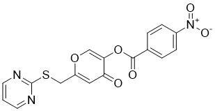
| SMILES Code | O=C(OC1=COC(CSC2=NC=CC=N2)=CC1=O)C3=CC=C([N+]([O-])=O)C=C3 |
| Synonyms | ML221; ML-221. |
| In Vitro | In vitro activity: Cells (angiotensin II receptor-like 1 (AGTRL-1) cell line (DiscoveRx, Cat# 93-0250C2)) were seeded at 1000 cell/well (1536 plate, Corning) in 4 μL and grown overnight (16-18 h) at 37 °C, 5% CO2, 100% humidity, then 60 nL of either DMSO control or 2 mM stock test compounds in DMSO were transferred to each well, followed by 2 μL of 30 nM Apelin-13 to negative control and test compound wells, and 2 μL of assay media (F12 nutrient mix HAMs supplemented with 10% hi-FBS, 1× penicillin/streptomycin) to positive control wells. This yielded a final concentration of test compound of 20 μM and 1% final DMSO. Assay was incubated for 90 min at room temperature, and then developed with 3 μL of detection reagent (PathHunter Detection Reagents (DiscoveRx, Cat# 93-0001)) for 60 min and luminescence read on a Perkin Elmer ViewLux. Kinase Assay: Antagonism of apelin-13-mediated activation of APJ by ML221 was assessed using two complimentary assays of APJ function; inhibition of cAMP and recruitment of β-arrestin. Increasing concentrations of ML221 antagonized a fixed concentration of Ap13 (EC80 = 10 nM) in both assays, with a calculated IC50equal to 0.70 μM in the cAMP assay, and 1.75 μM in the β-arrestin assay. Cell Assay: Apelin levels measured from supernatant (incubated for 48 h at 37 °C) from H69 and selected CCA cell lines using the Apelin-36 (human) EIA Kit according to the manufacturer’s instructions (Phoenix Pharmaceuticals, INC.). Undiluted samples (50 mL) were prepared in triplicates according to the protocol. Absorbance O.D. was measured at 450 nm on a microplate spectrophotometer (VersaMax, Molecular Devices, Sunnyvale, CA). The PRISM(R) software (GraphPad) was used to prepare the standard curve and to calculate the concentration of apelin in each sample. Data is expressed as an average concentration ± SEM. |
|---|---|
| In Vivo | Male BALB/c eight week old nude (nu/nu) mice were kept in a temperature and light controlled environment with free access to drinking water and rodent chow. Three million Mz-ChA-1 cells were suspended in extracellular matrix gel and subcutaneously injected into the rear flanks of these nude mice. Mice were treated with ML221 (150 μg/kg) 3× weekly via tail vein injection for 4 weeks. Tumor growth was measured three times a week using an electronic caliper, and volume was determined as follows: tumor volume (mm3) = length (mm) × width (mm) × height (mm). Tumors were allowed to grow until the maximum allowable tumor burden was reached, as set forth by the Baylor Scott & White Healthcare IACUC tumor burden policy. After 4 weeks of treatment, mice were euthanized with sodium pentobarbital (50 mg/kg i.p.). Hematoxylin and eosin (H&E) staining was performed using an H&E stain kit purchased from ScyTek Laboratories, INC. Tumors were confirmed to be primarily CCA cells by positive IHC staining and immunoblots for cytokeratin-19 (CK-19), a cholangiocyte specific marker. IHC and immunoblots were used to demonstrate expression of APLNR, p-ERK and t-ERK. Alpha tubulin was used as a relative control using a mouse monoclonal anti-alpha tubulin antibody purchased from abcam. Markers of proliferation (PCNA, Ki-67), angiogenesis (VEGF-A, VEGF-C, Ang-1, and Ang-2) and tumor progression (Vimentin, MMP-9, MMP-3) (Qiagen) were measured via rtPCR using the aforementioned protocol. |
| Animal model | Male BALB/c eight week old nude (nu/nu) mice |
| Formulation & Dosage | 150 μg/kg; 3× weekly via tail vein injection for 4 weeks |
| References | Bioorg Med Chem Lett. 2012 Nov 1; 22(21): 6656–6660. ; Cancer Lett. 2017 Feb 1;386:179-188. |
 m.cnreagent.com