| CAS NO: | 702674-56-4 |
| 规格: | ≥98% |
| 包装 | 价格(元) |
| 5mg | 电议 |
| 25mg | 电议 |
| 50mg | 电议 |
| 100mg | 电议 |
| 250mg | 电议 |
| 500mg | 电议 |
Molecular Weight (MW) | 471.35 |
Formula | C20H23BrN8O |
CAS No. | 702674-56-4 |
Storage | -20℃ for 3 years in powder form |
-80℃ for 2 years in solvent | |
Solubility (In vitro) | DMSO: 94 mg/mL (199.4 mM) |
Water: <1 mg/mL | |
Ethanol: N/A | |
Solubility (In vivo) | 30% PEG400+0.5% Tween80+5% propylene glycol: 30mg/mL |
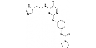
Synonyms | BX 912; BX912; BX912 Chemical Name: N-(3-((4-((2-(1H-imidazol-4-yl)ethyl)amino)-5-bromopyrimidin-2-yl)amino)phenyl)pyrrolidine-1-carboxamide InChi Key: DMMILYKXNCVKOJ-UHFFFAOYSA-N InChi Code: InChI=1S/C20H23BrN8O/c21-17-12-24-19(28-18(17)23-7-6-16-11-22-13-25-16)26-14-4-3-5-15(10-14)27-20(30)29-8-1-2-9-29/h3-5,10-13H,1-2,6-9H2,(H,22,25)(H,27,30)(H2,23,24,26,28) SMILES Code: O=C(N1CCCC1)NC2=CC=CC(NC3=NC=C(Br)C(NCCC4=CNC=N4)=N3)=C2 |
In Vitro | In vitro activity: BX912 prevents ChcK1, PKA, c-kit, and KDR with IC50 of 0.83, 0.11, 0.85, and 0.41 μM, resepectively. BX912 blocks PDK1/Akt signaling in tumor cells and suppresses the anchorage-dependent growth of a variety of tumor cell lines (such as PC-3 cells ) in culture or induces apoptosis. A number of cancer cell lines (such as MDA-468 breast cancer) with elevated Akt activity are>30-fold more sensitive to growth inhibition by PDK1 inhibitor BX912 in soft agar than on tissue culture plastic, consistent with the cell survival function of the PDK1/Akt signaling pathway, which is particularly important for unattached cells. BX912 potently blocks PDK1 enzyme activity in a direct kinase assay format, although BX912 fails to block preactivated AKT2 activity (IC50> 10 μM). Therefore, BX-912 is a direct inhibitor of PDK1. BX912 is a competitive inhibitor of PDK1 activity with respect to its substrate, ATP, suggesting that BX912 binds to the ATP binding pocket of PDK1. The aminopyrimidine backbone of BX912 adopts a similar orientation in the active site of PDK1. BX912 promotes a pronounced increase in the population of MDA-468 cells with 4 N DNA content, indicative of a block at the G2/M phase of the cell cycle. BX912 also potently inhibits the growth of HCT-116 cells in soft agar, showing a 96% inhibitory effect at a dose of 1 μM. BX912 potently inhibits the growth of PC-3 cells in soft agar, displaying IC50 of 0.32 μM.
Kinase Assay: PDK1 is assayed in a direct kinase assay and a coupled assay format measuring PDK1- and PtdIns-3,4-P2-mediated activation of AKT2. For the coupled assay, the final assay mixture (60 μL) contained: 15 mM MOPS, pH 7.2, 1 mg/mL bovine serum albumin, 18 mM β-glycerol phosphate, 0.7 mM dithiothreitol, 3 mM EGTA, 10 mM MgOAc, 7.5 μM ATP, 0.2 μCi of [γ- 33P]ATP, 7.5 μM biotinylated peptide substrate (biotin-ARRRDGGGAQPFRPRAATF), 0.5 μL of PtdIns-3,4-P2-containing phospholipid vesicles, 60 pg of purified recombinant human PDK1, and 172 ng of purified recombinant human AKT2. After incubation for 2 hours at room temperature, the biotin-labeled peptide is captured from 10 μL of the assay mixture on streptavidin-coated SPA beads, and product formation is measured by scintillation proximity in a Wallac MicroBeta counter. The product formed is proportional to the time of incubation and to the amount of PDK1 and inactive AKT2 added. PDK1 is added at suboptimal levels so that the assay could sensitively detect inhibitors of AKT2 activation as well as direct inhibitor BX912 of PDK1 or AKT2. To measure PDK1 activity directly, the final assay mixture (60 μL) contained 50 mM Tris-HCl, pH 7.5, 0.1 mM EGTA, 0.1 mM EDTA, 0.1% β-mercaptoethanol, 1 mg/mL bovine serum albumin, 10 mM MgOAc, 10 μM ATP, 0.2 μCi of [γ-33P]ATP, 7.5 μM substrate peptide (H2N-ARRRGVTTKTFCGT), and 60 ng of purified recombinant human PDK1. After 4 hours at room temperature, 25 mM EDTA is added and a portion of the reaction mixture on P81 phosphocellulose paper is spotted. The filter paper is washed three times with 0.75% phosphoric acid and once with acetone. After drying, the filter-bound labeled peptide is quantified using a phosphorimager.
Cell Assay: Cells such as MDA-468, MDA-453 are seeded at a low density (1.5-3 × 10³ cells/well, 0.1 mL/well, 96-well plates) and are incubated overnight. BX912 treatments are made by adding 10 μL/well of the compound in 1% dimethyl sulfoxide and growth medium (final concentration of dimethyl sulfoxide, 0.1%), followed by brief shaking. Treated cells are incubated for 72 hours, and viability is measured by the addition of 10 μL of the metabolic dye WST-1. The WST-1 signal is read in a plate reader at 450 nm, and a no cell, or zero time cell, background is subtracted to calculate the net signal. |
In Vivo | NA |
Animal model | NA |
Formulation & Dosage | NA |
References | J Biol Chem. 2005 May 20;280(20):19867-74. |
 m.cnreagent.com