| CAS NO: | 1271738-59-0 |
| 规格: | ≥98% |
| 包装 | 价格(元) |
| 25mg | 电议 |
| 50mg | 电议 |
| 100mg | 电议 |
| 250mg | 电议 |
| 500mg | 电议 |
| Molecular Weight (MW) | 375.55 |
|---|---|
| Formula | C18H25N5S2 |
| CAS No. | 1271738-59-0 |
| Storage | -20℃ for 3 years in powder form |
| -80℃ for 2 years in solvent | |
| Solubility (In vitro) | DMSO: 19 mg/mL (50.6 mM) |
| Water:<1 mg/mL | |
| Ethanol: 19 mg/mL (50.6 mM) | |
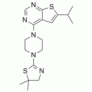
| Other info | Chemical Name: 4-[4-(5,5-dimethyl-4H-1,3-thiazol-2-yl)piperazin-1-yl]-6-propan-2-ylthieno[2,3-d]pyrimidine InChi Key: FUGQNAUKABUDQI-UHFFFAOYSA-N InChi Code: InChI=1S/C18H25N5S2/c1-12(2)14-9-13-15(20-11-21-16(13)24-14)22-5-7-23(8-6-22)17-19-10-18(3,4)25-17/h9,11-12H,5-8,10H2,1-4H3 SMILES Code: CC(C1=CC2=C(N3CCN(C4=NCC(C)(C)S4)CC3)N=CN=C2S1)C |
| Synonyms | MI 3; MI-3; MI3 |
| In Vitro | In vitro activity: In HEK293 cells, MI-3 accesses the protein target and effectively inhibits the menin-MLL-AF9 interaction. MI-3 effectively blocks MLL fusion protein-mediated leukemic transformation by downregulating the expression of target genes required for MLL fusion protein oncogenic activity. In human MLL leukemia cell lines harboring different MLL translocations, MI-3 effectively blocks cell proliferation, and induces apoptosis. Kinase Assay: FITC-MBM1 at 15 nM and menin at 150 nM in the FP buffer are mixed and incubated for 1h in the dark at room temperature. For point screening, the 0.2 μL of each compound (20 μM final concentration, 1% DMSO) is added to 20 μL of the aliquot of the protein-peptide mixture and incubated on 384-well plates in the dark at room temperature for 1h. In confirmation screening, the serial dilution plates with compounds in DMSO are prepared and used to titrate the menin-FITC-MBM1 complex. Change in fluorescence polarization is monitored at 525 nm after excitations at 495 nm using the PHERAstar microplate reader (BMG) and applied to determine IC50 values with the Origin 7.0 program. Cell Assay: The MLL-AF9 and E2A-HLF transduced murine BMC are plated in 12-well plates at the concentration of 5×10³ cells/mL with 1 mL methylcellulose medium M3234 containing 20% IMDM medium, 1% penicillin/streptomycin, IL-3 and 0.25% DMSO or compounds. 6 days later colonies are stained with 100 μL iodonitrotetrazolium chloride at final concentration of 1 mg/mL, incubated at 37°C for 30 min and counted. To replate for the 2nd round, colonies are counted at day 6 without staining and cells were washed out by 1×PBS buffer and resuspended in IMDM medium containing 15% FBS, 1% penicillin/streptomycin and IL-3. 5×10³ cells are plated in 12-well plates with 1ml methylcellulose medium M3234 containing 20% IMDM medium, 1% penicillin/streptomycin, IL-3 and 0.25% DMSO or compounds. 6 days later colonies are stained and counted. |
|---|---|
| In Vivo | MLL-AF9 transformed BMC that remained viable after 7 days of treatment with MI-2 and MI-3 showed substantial changes in morphology, indicative of monocytic differentiation, as evidenced by increased cell size, lower nuclear to cytoplasmic ratio and highly vacuolated cytoplasm. Consistent with the change in cell morphology, the expression of CD11b was substantially increased on MLL-AF9 transformed BMC after 7 days of treatment with MI-2 and MI-3. |
| Animal model | NA |
| Formulation & Dosage | NA |
| References | Nat Chem Biol. 2012 Jan 29;8(3):277-84. |
 m.cnreagent.com