您好,欢迎来到化工原料网! [登录] [免费注册]
化工原料网
位置:首页 > 产品库 > CY5
立即咨询
咨询类型:
     
*姓名:
*电话:
*单位:
Email:
*留言内容:
请详细说明您的需求。
*验证码:
 
CY5
本产品不向个人销售,仅用作科学研究,不用于任何人体实验及非科研性质的动物实验。
CY5图片
CAS NO:146368-11-8
包装与价格:
包装价格(元)
10 mM * 1 mL in DMSO电议
5mg电议
10mg电议
25mg电议
50mg电议
100mg电议
200mg电议

产品名称
Sulfo-Cyanine5
产品介绍
CY5 是一种 CY 染料。CY 为花菁 (Cyanine) 的缩写,是由奇数个甲基单位连接的两个氮原子组成的化合物。菁类化合物具有波长长、吸收和发射可调、消光系数高、水溶性好、合成相对简单等特点。CY 系类染料常被用于蛋白,抗体以及小分子化合物的标记,对于蛋白抗体的标记,可以通过简单的混合反应来完成结合,以下我们介绍了蛋白抗体标记的标记方法,具有一定的参考意义。
生物活性

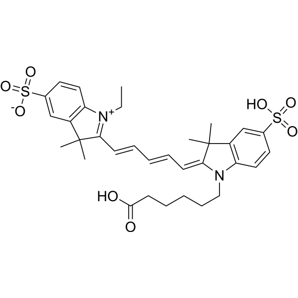
CY5 is a CY dye. CY, short for Cyanine, is a compound consisting of two nitrogen atoms connected by an odd number of methyl units. Cyanine compounds have the characteristics of long wavelength, adjustable absorption and emission, high extinction coefficient, good water solubility and relatively simple synthesis[1]. CY dyes are of en used for the labeling of proteins, antibodies and small molecular compounds. For the labeling of protein antibodies, the combination can be completed through a simple mixing reaction. Below, we introduce the labeling method of protein antibody labeling, which has certain reference significance[2].

体外研究
(In Vitro)

原液制备
1. 蛋白准备
为了获得最佳标记效果,请将蛋白 (抗体) 浓度配制为 2 mg/mL。
1) 蛋白溶液 pH 值应为 8.5±0.5,若 pH 低于 8.0,则用 1 M 碳酸氢钠进行调整。
2) 若蛋白浓度低于 2 mg/mL,标记效率会大大降低,为了获得最佳标记效率,建议最终蛋白质浓度范围为 2-10 mg/mL。
3) 蛋白必须在不含伯胺 (如 Tris 或甘氨酸) 和铵离子的缓冲液中,否则会影响标记效率。
2. 染料准备
将无水 DMSO 稀释 CY 染料,制成 10 mM 储备溶液。通过玻璃管或旋涡充分混合。
注:CY 储存液建议分装后于 -20 ℃ 或 -80 ℃ 避光保存。
3. 染料工作液用量计算
标记反应所需的 CY 染料用量取决于要标记蛋白的用量,CY 染料与蛋白的最佳摩尔比为 10 左右。
例:假如所需标记蛋白为 500 μL 2 mg/mL 的 IgG (MW=150,000),用 100 μL DMSO 溶解一管 1 mg CY 染料,则所需 CY 体积为 3.95 μL,详细计算流程如下 (以 CY3-NHS ester 为例) :
1) mmol (IgG) = mg/mL (IgG) ×mL (IgG) / MW (IgG) =2 mg/mL×0.5 mL / 150,000 mg/mmol= 6.7×10⁻⁶ mmol
2) mmol (CY3-NHS ester) = mmol (IgG) ×10 =6.7×10⁻⁶ mmol×10 = 6.7×10⁻⁵ mmol
3) μL (CY3-NHS ester) = mmol (CY3-NHS ester) ×MW (CY3-NHS ester) / mg/μL (CY3-NHS ester) = 6.7×10⁻⁵ mmol× 590.15 mg/mmol / 0.01 mg/μL =3.95 μL (CY3-NHS ester)


使用方法
1. 标记反应
1) 取算好体积的新鲜配制的 10 mg/mL CY 染料缓慢加入到 0.5 mL 蛋白样品溶液中,轻轻摇匀混合,然后短暂离心将样品收集在反应管底部。切忌剧烈混匀,防止蛋白样品变性失活。
2) 将该反应小管置于避光处,在室温条件下轻轻摇晃孵育 60 分钟,每隔 10–15 分钟,将反应小管轻轻颠倒几次,以充分混合两种反应物,提高标记效率。
2. 蛋白纯化脱盐
以下方案是使用 SepHadex G-25 柱纯化染料蛋白偶联物为例。
1) 按照生产说明书制备 SepHadex G-25 柱。
2) 将反应混合物装入 SepHadex G-25 色谱柱顶部。
3) 当样品运行到顶部树脂表面下方时,立即加入 PBS (pH 7.2-7.4)。
4) 向所需样品中加入更多的 PBS (pH 7.2-7.4),完成柱纯化。结合含有所需要的染料-蛋白质缀合物的组分。

分子量

656.81

性状

Solid

Formula

C33H40N2O8S2

CAS 号

146368-11-8

Emission (Em)
Em 620-750 nm Red
Excitation (Ex)
Ex 620-750 nm Red
运输条件

Room temperature in continental US; may vary elsewhere.

储存方式

4°C, protect from light

*In solvent : -80°C, 6 months; -20°C, 1 month (protect from light)

溶解性数据
In Vitro: 

DMSO : 125 mg/mL(190.31 mM;Need ultrasonic)

H2O : 25 mg/mL(38.06 mM;Need ultrasonic)

配制储备液
浓度溶剂体积质量1 mg5 mg10 mg
1 mM1.5225 mL7.6126 mL15.2251 mL
5 mM0.3045 mL1.5225 mL3.0450 mL
10 mM0.1523 mL0.7613 mL1.5225 mL
*

请根据产品在不同溶剂中的溶解度选择合适的溶剂配制储备液;一旦配成溶液,请分装保存,避免反复冻融造成的产品失效
储备液的保存方式和期限:-80℃, 6 months; -20℃, 1 month (protect from light)。-80℃ 储存时,请在 6 个月内使用,-20℃ 储存时,请在 1 个月内使用。

In Vivo:

请根据您的实验动物和给药方式选择适当的溶解方案。以下溶解方案都请先按照In Vitro方式配制澄清的储备液,再依次添加助溶剂:

——为保证实验结果的可靠性,澄清的储备液可以根据储存条件,适当保存;体内实验的工作液,建议您现用现配,当天使用; 以下溶剂前显示的百
分比是指该溶剂在您配制终溶液中的体积占比;如在配制过程中出现沉淀、析出现象,可以通过加热和/或超声的方式助溶

  • 1.

    请依序添加每种溶剂: 10% DMSO    40%PEG300   5%Tween-80   45% saline

    Solubility: ≥ 2.08 mg/mL (3.17 mM); Clear solution

    此方案可获得 ≥ 2.08 mg/mL (3.17 mM,饱和度未知) 的澄清溶液。

    以 1 mL 工作液为例,取 100 μL 20.8 mg/mL 的澄清 DMSO 储备液加到 400 μL PEG300 中,混合均匀;向上述体系中加入50 μL Tween-80,混合均匀;然后继续加入 450 μL生理盐水定容至 1 mL。

    将 0.9 g 氯化钠,完全溶解于 100 mL ddH2O 中,得到澄清透明的生理盐水溶液
  • 2.

    请依序添加每种溶剂: 10% DMSO    90% (20%SBE-β-CDin saline)

    Solubility: ≥ 2.08 mg/mL (3.17 mM); Clear solution

    此方案可获得 ≥ 2.08 mg/mL (3.17 mM,饱和度未知) 的澄清溶液。

    以 1 mL 工作液为例,取 100 μL 20.8 mg/mL 的澄清 DMSO 储备液加到 900 μL20% 的 SBE-β-CD 生理盐水水溶液中,混合均匀。

    将 2 g 磺丁基醚 β-环糊精加入 5 mL 生理盐水中,再用生理盐水定容至 10 mL,完全溶解,澄清透明
*以上所有助溶剂都可在本网站选购。
染色示例
  • Description: CY5 is a far-red-fluorescent hydrophobiccan for label peptides, proteins and oligonucleotides.
    Method: For immunofluorescence assay.
    1. The testing protein used in immunofluorescence is labeled by Cy5 dye.
    2. Immunofluorescence images are acquired by Zeiss 689 LSM 980 microscope (Zeiss, Germany) with ZEN Connect software.
  • Description: CY5 is a far-red-fluorescent hydrophobiccan for label peptides, proteins and oligonucleotides.
    Method: For nanoparticles labeling.
    1. Cy5 (2.0 mg/mL) methanol solution is prepared as a pre-solution, and 10 mL of the pre-solution is added slowly to 10 mL of selenium nanoparticles with continuous stirring at 300 rpm for 6 h at room temperature. All operations are carried out in the dark.
    2. The unreacted Cy5 is removed by dialysis, and continuous dialysis is carried out until no fluorescence is detected in the dialysate.
    3. The product is collected to obtain selenium nanoparticles Cy5. For preparing chitosan-Cy5, a chitosan solution with a concentration equal to that of chitosan in selenium nanoparticles is used to replace selenium nanoparticles to obtain chitosan-Cy5.
  • Description: CY5 is a far-red-fluorescent hydrophobiccan for label peptides, proteins and oligonucleotides.
    Method: For determine the localization of cubosomes.
    1. Co-dissolve Cy5 (2% w/w) with the lipid mixtures in chloroform, the Cy5 labeled cubosomes are loaded in dialysis cassettes to remove any free dye.
    2. Thirty xenografts mice are divided into two equal group sizes with 15 animals in each. One of the groups received Cbs-Cy5 and the second group Cbs-Cy5-Af (Affimer targeted). The administration is done through the tail vein of the mice with 100 μL of sample, equating to 50 mg/kg of cubosome to mouse body weight.
    3. Localization of the Cy5-Cb in the mice is studied by using the Cy5 fluorescence filters in the IVIS Spectrum for a duration of up to 72 h postinjection. At each time point, three mice are euthanized, and the brain, liver, kidney, spleen, heart, and lungs along with the tumor are scanned under the IVIS to quantify the Cy5 fluorescence. The tissues are then frozen in OCT. Sections of 5 μm thickness are made by using a cryostat (Leica CM3050S) and are examined under a confocal microscope (Nikon A1R LSM).