| 包装 | 价格(元) |
| 10mM (in 1mL DMSO) | 电议 |
| 100mg | 电议 |
Cell experiment: | 1% aqueous solution of Nile blue A is prepared and filtered before use. Mild heating may be necessary to fully dissolve the stain. Heat-fixed smears of bacterial cells are stained with the Nile blue A solution at 55℃ for 10 min in a coplin staining jar. After being stained, the slides are washed with tap water to remove excess stain and with 8% aqueous acetic acid for 1 min. The stained smear is washed and blotted dry with bibulous paper, remoistened with tap water, and covered with a no. 1 glass cover slip. The preparation is examined with a Nikon Labphot microscope with an episcopic fluorescence attachment[1]. The PHA– strain Escherichia coli UT5600(DE3) is stained with Nile blue A. In an Erlenmeyer flask, 20 mL of Luria–Bertani (LB) broth is inoculated with one colony of UT5600(DE3) and incubated for 14 h at 37 ℃ and 200 rpm. Subsequently, 20 mL of LB broth containing Nile blue A in a final concentration of 0.5 μg/mL is inoculated with 200 μL of the 14-h culture and cultured to an optical density at 578 nm (OD578) of 0.6. As a control, 20 mL of LB broth (without Nile blue A) is inoculated with 200 μL of the 14-h culture and is also cultured to an optical density at OD578 of 0.6. Every 20 min, the OD578 is determined for both cultures to verify whether there is any influence of the dye on the growth of the bacteria[2]. Nile blue A stock solution is prepared using ethanol as the solvent. The concentration of NB is maintained at 5 μM for all the studies. The solutions are left for 1 h to achieve equilibrium before spectral measurements. The absorption spectra are recorded using Shimadzu Spectrophotometer (UV-1800) and the emission spectra are recorded using Jobin–Yvon Spectrofluorimeter. A 450 nm nano-LED is used as the light source and the fluorescence lifetime is collected at λem=672 nm[3]. |
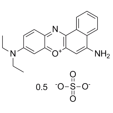
| 产品描述 | Nile Blue A is used to differentiate melanins and lipofuscins. It is also useful for staining fats and preparation of an amperometric glucose sensor. Nile blue A is a basic oxazine dye which is soluble in water and ethyl alcohol. Nile blue A is a satisfactory stain for PHB granules in bacteria and is in fact superior to Sudan black B for this purpose. Poly-p3-hydroxybutyrate granules exhibits a strong orange fluorescence when stained with Nile blue A. Nile blue A appears to stain many more PHB granules than Sudan black B does and is not as easily ished from the cell by decolorization procedures[1]. Nile blue A is used as a stain for polyhydroxyalkanoic acid-accumulating microorganisms or to detect polyhydroxyalkanoic acids in microorganisms. Escherichia coli cells that do not accumulate detectable polyhydroxyalkanoic acids can be stained with Nile blue A and that this staining is sufficient for identifying these cells in fluorescence-activated cell sorting (FACS) experiments. Nile blue A staining does not affect either surface display of peptides or specific labeling of these peptides by a second fluorescence. Staining E. coli for flow cytometry using Nile blue A is an easy-to-handle and low-cost alternative to other fluorescent dyes or the intracellular expression of, for example, green fluorescent protein[2]. Nile blue A is one of the most studied benzophenoxazine dyes, as a potent photosensitizer for photodynamic therapy. The dye when administered intravenously disperses throughout the body by circulating through blood and is taken up by most cells that emphasize its interaction with various biomolecule[3]. [1]. Ostle AG, et al. Nile blue A as a fluorescent stain for poly-beta-hydroxybutyrate. Appl Environ Microbiol. 1982 Jul;44(1):238-41. [2]. Betscheider D, et al. Nile blue A for staining Escherichia coli in flow cytometer experiments. Anal Biochem. 2009 Jan 1;384(1):194-6. [3]. Mishra SS, et al. Spectroscopic investigation of interaction of Nile Blue A, a potent photosensitizer, with bile salts in aqueous medium. J Photochem Photobiol B. 2014 Dec;141:67-75. |
 m.cnreagent.com